eConsult Transcript
PCP submission
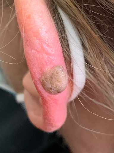
50-year old White adult female with PMH of HTN, every-day smoker, vitamin D deficiency, RA with multiple joints pain and depression presents to office visit for unexpected lesion on left earlobe appearing brown to dark color, asymmetric, 3-4cm in diameter, and mildly regular border. Needing recommendation.
Specialist response
Thank you for the consult. I favor this is most compatible with a seborrheic keratosis (SK). SKs are extremely common benign neoplasms of the epidermis that can occur on the head, neck, trunk, and extremities. SKs tend to increase in incidence and number with increasing age, and as in this case are characterized by a waxy, stuck-on or sometimes verrucous appearance. They are generally removed only for cosmetic reasons, though treatment with cryotherapy can be helpful if they become irritated or inflamed. Sometimes SKs can have overlapping morphologic features with melanocytic lesions and a biopsy may be prudent to distinguish the two. But in most cases, patient reassurance regarding the chronic and benign nature of these lesions is all that is needed. As such I recommend: — Patient education on the diagnosis of SK as above. — If the patient desires removal, I would either trial cryotherapy with liquid nitrogen (spray 3-5 secs x 2 cycles — let it thaw completely in between cycles) or do shave removal with tissue sent to pathology. Despite the clinical impression being of relatively high confidence, it is generally not recommended to do any kind of excisional biopsy without obtaining histologic confirmation because of the potential (albeit small) for morphologic overlap of some malignant cutaneous lesions. Those are my initial thoughts and I hope this helps. I am happy of course to discuss further. Thanks again for the case!

